


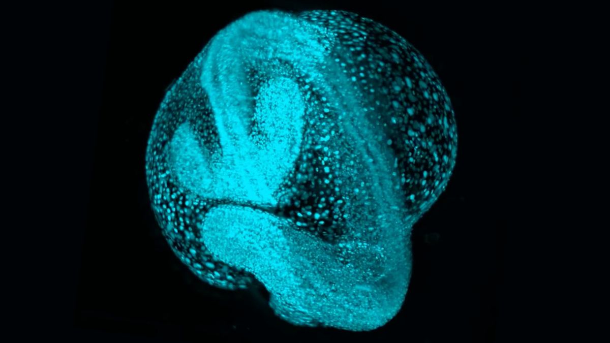
Striking new psychedelic videos give a glimpse into what living organisms look like during their earliest moments — and it took scientists years to capture.
The videos are part of a new atlas of embryos called Zebrahub, which shows where cells are located and what they’re doing at different stages of development. The atlas combines high-resolution timelapse videos of developing embryos with data revealing which genes are switched on at each developmental stage.
The atlas covers the embryos of zebrafish (Danio rerio), a type of minnow often used in biological research. The majority of the small fishes’ genes have close analogs in humans, and the major components of cells are common across the vertebrate branch of the tree of life.
“At these early stages of life, all embryos are very similar,” said Loïc Royer, one of the developers of Zebrahub, leader of the Organismal Architecture group and director of imaging AI at the Chan Zuckerberg Biohub San Francisco. “The shapes, the genes, the molecular machines that are responsible for doing that work of building an organism — it’s all very similar.”
Related: Early development is inherently ‘chaotic,’ new atlas of mammal embryos reveals
Royer is the senior author of a new paper describing Zebrahub, published Thursday (Oct. 24) in the journal Cell. He said that it’s difficult to predict what kind of discoveries the new tool might enable, but studying the embryos of other lifeforms could address questions about how birth defects and other congenital disorders arise in humans. In addition, the new atlas may hold clues to why animals like zebrafish can regenerate their body parts following an injury, but we cannot, he suggested. And it could reveal key differences between youthful and aging tissues, which could help explain why we grow old.
At its core, Zebrahub focuses on one central question. “It’s essentially the question of how we are built,” Royer told Live Science. “If we don’t know how we’re built, how do we hope to ‘repair’ ourselves?”
Zebrahub is free to access and offers tools to help biologists view and use the trove of data. To collect the data in the first place, though, Royer and colleagues needed to develop new methods of studying zebrafish embryos.
Historically, studies have focused on either where cells are located within a developing embryo or which genes are active at a given moment. To track cells’ locations, scientists take many snapshots of the embryos under a microscope. Zebrahub’s developers created a new microscope that sweeps a thin sheet of light across the whole embryo, generating pictures as it goes. This technique avoids exposing the embryos to harsh lasers that could harm them.
Image 1 of 4


The team used their microscope to capture timelapses of embryos from the time of fertilization through about 24 hours of growth. (Zebrafish hatch about three to four days after fertilization, so by Day 1, organs already start to form.) The researchers then analyzed these timelapses using a new software designed to track the movements of each individual cell in 3D space.
Historically, to track which genes in the embryo are switched on, researchers had to “melt” the embryos down, turning them into a “soup” that can then be analyzed by a machine, Royer explained. The problem is that you need 30 to 60 embryos, because turning them to soup inevitably damages some of their genetic material, limiting what’s left to analyze.
Related: ‘First complete models’ of a human embryo made in the lab
The Zebrahub developers found ways of handling embryos very gently, preserving them well enough to analyze just one embryo at a time. They looked at more than 120,400 cells from 40 zebrafish embryos and larvae that ranged from 10 hours to 10 days old. They sequenced all the cells’ RNA — a molecule that enables cells to make proteins from DNA’s blueprints. The identity of a given cell can then be discerned from its gene activity.
At this level of resolution, the scientists spotted types of cells that tend to be missed through other methods, Royer said. For instance, they identified special stem cells — called neuro-mesodermal progenitors — and showed that they transformed into both nerve cells and muscle cells over time. It had been thought that the cells only gave rise to nerves.
Currently, the data in Zebrahub is based on two sets of embryos: one for the timelapses and one for the RNA. Nonetheless, these datasets can be compared to give scientists an idea of what an embryo looks like as certain genes are switched on. Looking forward, Royer and colleagues are working on collecting the same kinds of information from a single set of embryos, to better marry the data.
In the meantime, other groups of scientists are already using Zebrahub as a starting point to study human conditions. For example, one group combined Zebrahub with their own cell data to probe which proteins might drive cataracts to form in the eyes. They were able to see when various genes switch on and off as the lens of the eye first develops.
“We study fish because we cannot study human embryos, for obvious reasons,” Royer said. “What we learn from the embryos, we learn about ourselves — so I study fish because I want to study myself.”
Ever wonder why some people build muscle more easily than others or why freckles come out in the sun? Send us your questions about how the human body works to community@livescience.com with the subject line “Health Desk Q,” and you may see your question answered on the website!


")


