Arguably the most complex organ in the body, the brain is an elegant mess of cells, chemicals and electrical impulses that orchestrates our thoughts, behaviors and unconscious bodily functions.
This year, Live Science covered a slew of fascinating studies about the brain, each of which revealed new insights about how the organ ticks while also raising new questions.
Related: Master regulator of inflammation found — and it’s in the brain stem
The male hormone cycle and the brain
Although not often discussed, the male hormone cycle is quite striking. Steroid hormones in the male body — including testosterone, cortisol and estradiol — decrease about 70% throughout the day and then reset overnight. This year, a brain-scan study revealed that the brain loses and regains volume in time with this daily cycle. At this point, though, it’s unclear whether the hormones themselves drive the brain changes or how this cycle affects male brain function.
Your brain on cinema

Many people throw on a movie when they want to “turn off their brain” for a bit. But a recent study found that, actually, 24 different brain networks light up as you watch different types of films. By tying brain activity patterns to what was happening in a given scene, the scientists behind the study were able to construct the most accurate functional brain map to date.
Transformation of babies’ brains after birth

A groundbreaking study in fetuses and newborns highlights how the activity of certain parts of the brain suddenly shift after birth. There is a huge uptick in activity in the subcortical network, which acts like a relay hub for information, and the sensorimotor network, which is responsible for processing external stimuli and coordinating movements. The researchers now want to study the same brain networks in preterm babies, to see if there’s a notable difference from full-term babies.
The drive to eat

A simple circuit made of just three types of neurons may underlie our drive to eat, a study found. The brain cells work together to detect hunger-signaling hormones and ultimately set off cells that control the muscles for chewing. By messing with this circuit in lab mice, the researchers prompted the rodents to eat 12 times more food than usual and make chewing movements even when they had no food.
Pregnancy’s “permanent etchings” on the brain
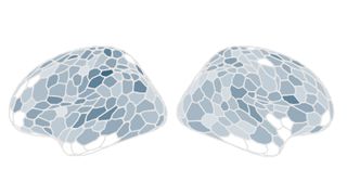
Throughout pregnancy, about 80% of the brain’s gray matter loses volume. Some of this lost volume reappears postpartum, but much of the missing matter stays lost. A similar loss of gray-matter volume is seen during puberty, a time when the brain is “pruning” away excess connections to boost its efficiency. It may be that this change in pregnancy reflects a similar fine-tuning of brain circuitry, scientists say.
Three copies of each memory
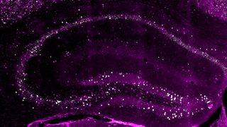
A mouse study suggests that the brain may store at least three copies of every memory it encodes. These copies differ in when they’re created, how long they last and how modifiable they are over time. Understanding when and how these different copies form could help scientists address conditions that affect memory encoding and retrieval, such as PTSD and dementia.
Conscious lab-grown brains?
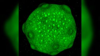
Could brain organoids — miniature models of the brain grown in the lab — ever gain consciousness? Live Science posed the question to neuroscientist Kenneth Kosik, who assured us that it’s not likely to happen anytime soon. However, there is still ongoing debate about what qualifies as “consciousness,” he noted. And there are big questions about what could happen if human minibrains were transplanted into animals or hooked up to technologies to create so-called cyborgs.
Related: Optical illusion reveals key brain rule that governs consciousness
‘Shrooms that “dissolve” sense of self

Psilocybin, the active ingredient in magic mushrooms, may cause the brain network responsible for maintaining a person’s sense of self to fall out of sync. Called the default mode network, this brain circuit is most active when people are self-reflecting and not engaged in any particular task. On high-dose psilocybin, the network’s activity temporarily changed, with some of the effects lasting a few weeks.
Origin of psychosis
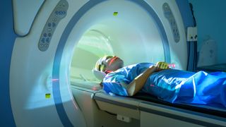
A brain-scan study could help confirm a theory about why people experience psychosis, or sudden breaks from reality. Using artificial intelligence (AI) to analyze the scans, scientists found overlapping “signatures” in the brains of people with psychosis. These signatures were either tied to a genetic condition or were from an unknown cause. The findings back a theory that, in psychosis, brain networks in charge of directing a person’s attention malfunction, which leads to hallucinations and delusions.
“Universal” brain pattern in primates

Multiple primate species, including humans, share a common brain-wave pattern in the cerebral cortex, the outer surface of the brain. This universal pattern is marked by high-frequency brain waves in the upper layers of the cortex and slower waves in its deeper layers. Scientists think the interplay of fast and slow waves dictates which information remains in conscious thought at any given moment.
Flow state
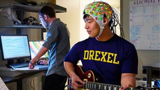
Scientists revealed the key parts of the brain that switch on when someone is in a flow state — or “in the zone.” The study involved scanning the brains of musicians with varying degrees of experience. It pitted two competing theories of “flow” against each other, and one theory came out on top.
Reading in a “blink”

A study found that the human brain can discern the basic structures of written language in the time it takes to blink. That suggests the brain can process written phrases about as quickly as it does visual scenes of the world around us. The study’s subjects seemed to process phrases that included subjects, verbs and objects faster than they did lists of nouns. They were also quick to process when the meaning of a phrase didn’t quite make sense. The findings suggest the brain isn’t only detecting that words are present but also starting to parse meaning right away.
Big brains from gut bugs
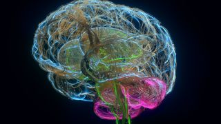
A mouse study suggests that humans’ remarkably big brains may have evolved partly thanks to the unique microbes in our guts. The study researchers transplanted microbes from the guts of humans and nonhuman primates into mice. The microbes from humans and big-brained primates converted food into energy for the brain more efficiently compared with the microbes from small-brained monkeys. The findings suggest that the big-brain microbes convert food into energy for the brain more efficiently, thus helping fuel the organ’s growth.
Stunning new brain map
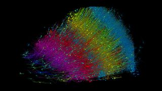
Researchers from Harvard and Google unveiled a striking map of a small sliver of the human brain. It contains roughly 57,000 neurons, 9 inches (23 centimeters) of blood vessels and 150 million synapses (the connection points between neurons). The map highlighted unique and unexpected features of the brain, including mysterious “whorls,” or knots, in the outgoing wires of some neurons.
3D-printed brain tissue that actually works
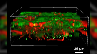
In a first, scientists 3D printed functional human brain tissue. The printer itself used stem cells instead of ink, and the team then used a concoction of chemicals to spur those cells to transform into brain cells. The resulting cells could communicate and link up in networks, similar to cells in an actual brain.
Ever wonder why some people build muscle more easily than others or why freckles come out in the sun? Send us your questions about how the human body works to [email protected] with the subject line “Health Desk Q,” and you may see your question answered on the website!


